Anatomy Of Musckes Sndctendons / Muscle and Tendon Characteristics - Classic Human Anatomy ... : Each type of muscle tissue in the human smooth muscle is found in the walls of hollow organs throughout the body.
byAdmin-
0
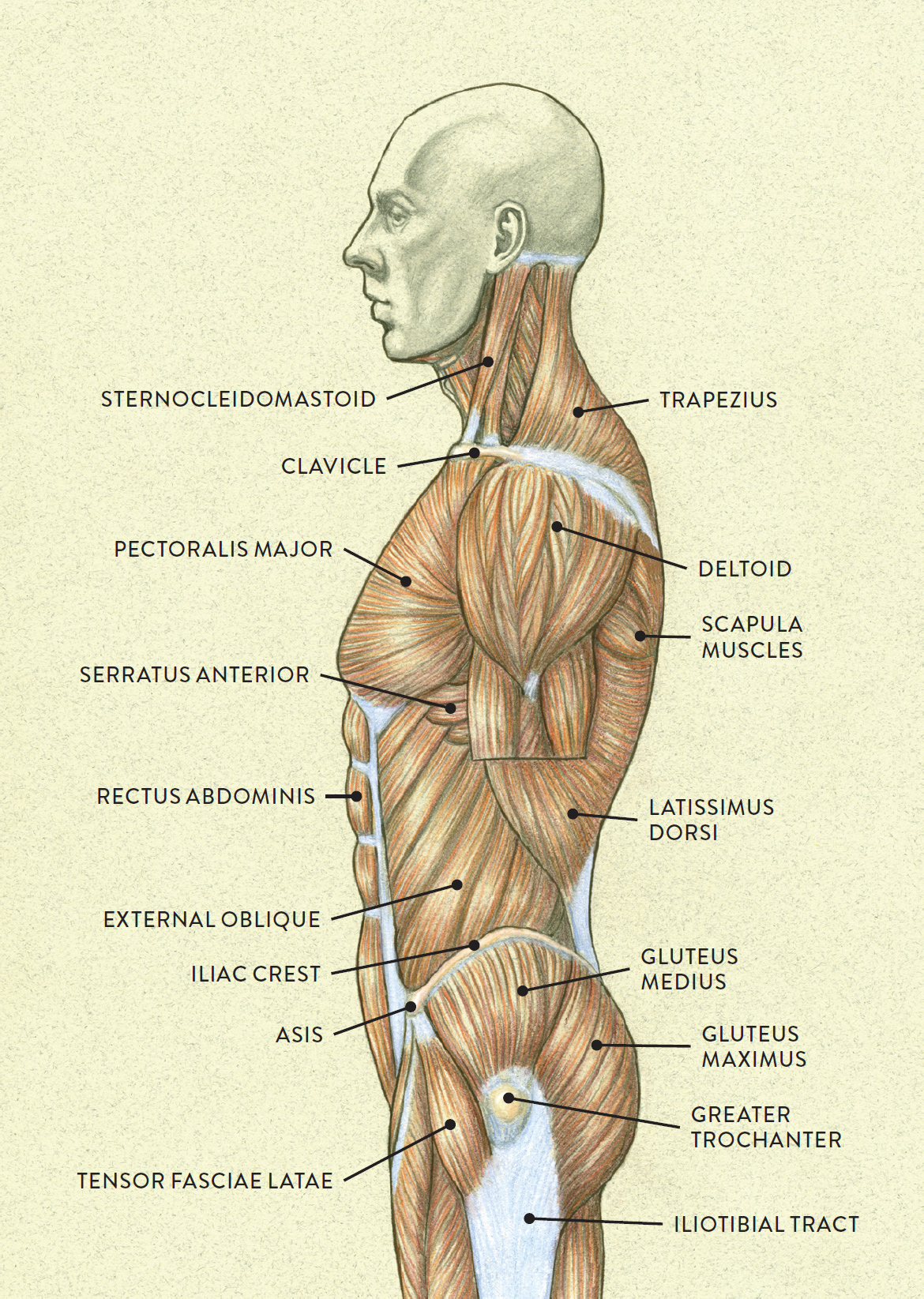
Anatomy Of Musckes Sndctendons / Muscle and Tendon Characteristics - Classic Human Anatomy ... : Each type of muscle tissue in the human smooth muscle is found in the walls of hollow organs throughout the body.. The interactive muscle anatomy diagram shown below outlines the major superficial (i.e. There's no strict demarcation or dividing line between the tendon and the covering around this muscle but that covering is called is called the epimysium fp my cm and it's really just connective tissue that covers the muscle kind of protects it reduces friction. Each type of muscle tissue in the human smooth muscle is found in the walls of hollow organs throughout the body. This article will focus on tongue embryology, origin, insertion, and action of the extrinsic muscles, followed by innervation, blood supply and lymphatic drainage of the tongue. There are around 650 skeletal muscles within the typical human body.
The text describes the concept of fascial continuum, which explains the. All about the shoulder muscles. Topographically, the muscles in this group are classed along with the lateral torso wall and upper extremity , which is due to their location as well as their genetic development based on their embryological origin. The interactive muscle anatomy diagram shown below outlines the major superficial (i.e. There are around 650 skeletal muscles within the typical human body.
MUSCLES OF THE TORSO INDICATED BY COLOR from schoolbag.info The muscles of mastication are a group of muscles associated with movements of the jaw. Muscular contraction is necessary for voluntary and involuntary movement of limbs, stabilization of joints, maintaining luminal diameter (in the case of arteries, bowel, etc), and to produce heat. The muscular system is responsible for the movement of the human body. But muscle is also the dominant tissue in the heart and in the walls of other hollow organs of the body. What of anatomy an essential textbook. The broad muscle that covers the top of the skull raises eyebrows, wrinkles forehead In the muscular system, muscle tissue is categorized into three distinct types: This article will focus on tongue embryology, origin, insertion, and action of the extrinsic muscles, followed by innervation, blood supply and lymphatic drainage of the tongue.
The text describes the concept of fascial continuum, which explains the.
By contracting, they also aid the venous return of blood to the heart and with age, these components of the musculoskeletal system progressively degenerate, which contributes to frailty and increases the risk of falls and fractures. The muscular system is responsible for the movement of the human body. There's no strict demarcation or dividing line between the tendon and the covering around this muscle but that covering is called is called the epimysium fp my cm and it's really just connective tissue that covers the muscle kind of protects it reduces friction. Digastric, mylohyoid, geniohyoid, stylohyoid infrahyoid muscles: How to study muscle anatomy. Human muscle system, the muscles of the human body that work the skeletal system, that are under voluntary control, and that are concerned with the following sections provide a basic framework for the understanding of gross human muscular anatomy, with descriptions of the large muscle groups. What of anatomy an essential textbook. The muscles of mastication develop from the first pharyngeal arch. However, if you take a little time to learn a few root words, those latin names can give you valuable insights into things like the muscle's size and shape. The text describes the concept of fascial continuum, which explains the. Inflammation of this region caused by repetitive stress or trauma may lead to pain and numbness known as carpal tunnel syndrome. This article will focus on tongue embryology, origin, insertion, and action of the extrinsic muscles, followed by innervation, blood supply and lymphatic drainage of the tongue. Home > blog > anatomy > shoulder anatomy:
Topographically, the muscles in this group are classed along with the lateral torso wall and upper extremity , which is due to their location as well as their genetic development based on their embryological origin. It elevates and protrudes the mandible. Roll your mouse over any muscle in the diagram below to learn its name. Their main function is contractibility. The broad muscle that covers the top of the skull raises eyebrows, wrinkles forehead
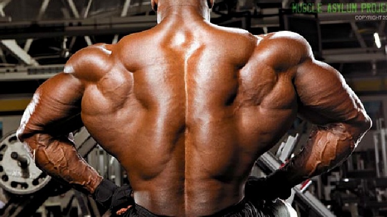
Deep Muscles of the Back - Erector Spinae • Bodybuilding ... from bodybuilding-wizard.com There's no strict demarcation or dividing line between the tendon and the covering around this muscle but that covering is called is called the epimysium fp my cm and it's really just connective tissue that covers the muscle kind of protects it reduces friction. Knee function is determined in large part by the anatomy of the joint. This is a table of skeletal muscles of the human anatomy. Inflammation of this region caused by repetitive stress or trauma may lead to pain and numbness known as carpal tunnel syndrome. Lesson on the anatomy of the forearm: What of anatomy an essential textbook. Tendons are tough bands of dense. Rectus capitis, longus capitis, longus colli.
Convergent muscles contain fibers that have a wide origin, but converge in order to attach to a narrow tendon.
What of anatomy an essential textbook. A collection of anatomy notes covering the key anatomy concepts that medical students need to learn. Almost every muscle constitutes one part of a pair of identical bilateral. The muscles of mastication are a group of muscles associated with movements of the jaw. The broad muscle that covers the top of the skull raises eyebrows, wrinkles forehead All about the shoulder muscles. The muscles of the torso, examined in the previous chapter, include a few that attach directly into the upper arm and help move the humerus at the shoulder joint. Rectus capitis, longus capitis, longus colli. Located immediately below the skin) muscles of the body. Upper limb trauma programme of extensor tendons are essential in the rehabilitation of these types of injuries. Lesson on the anatomy of the forearm: Digastric, mylohyoid, geniohyoid, stylohyoid infrahyoid muscles: Along with lateral pterygoid muscle it produces side to side movement of mandible.
In all its forms, it makes up nearly half of the body's mass. There's no strict demarcation or dividing line between the tendon and the covering around this muscle but that covering is called is called the epimysium fp my cm and it's really just connective tissue that covers the muscle kind of protects it reduces friction. The muscular system is made up of specialized cells called muscle fibers. Home > blog > anatomy > shoulder anatomy: Tendons are tough bands of dense.
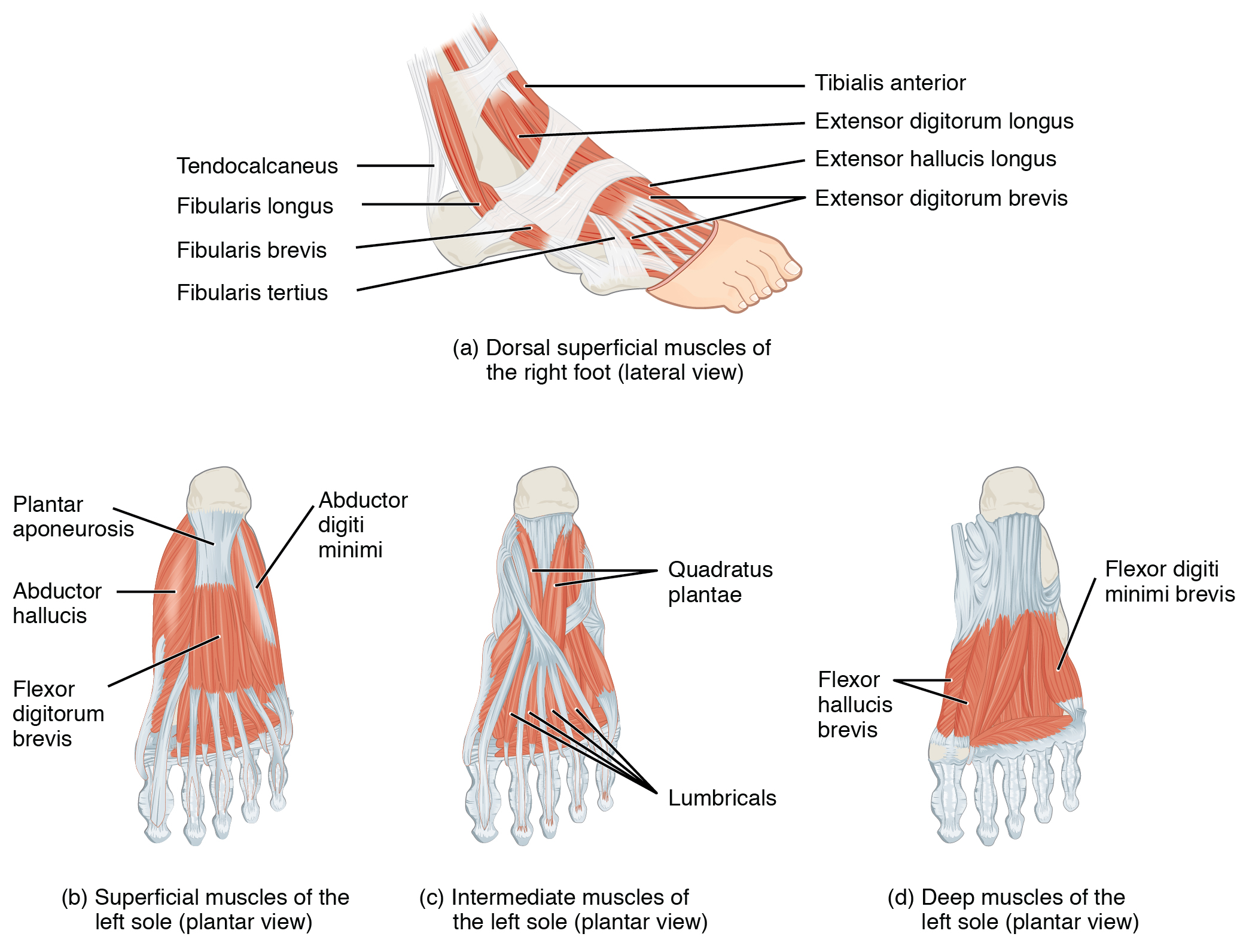
Appendicular Muscles of the Pelvic Girdle and Lower Limbs ... from philschatz.com Lesson on the anatomy of the forearm: Home > blog > anatomy > shoulder anatomy: You can click on any highlighted muscle to view a more detailed image of the. Sternohyoid, sternothyroid, thyrohyoid, omohyoid anterior vertebral muscles: In all its forms, it makes up nearly half of the body's mass. This review will also illustrate the vascular and lymphatic network and the innervating nerve branch. The broad muscle that covers the top of the skull raises eyebrows, wrinkles forehead Learn anatomy faster and remember everything you learn.
This review will also illustrate the vascular and lymphatic network and the innervating nerve branch.
Inflammation of this region caused by repetitive stress or trauma may lead to pain and numbness known as carpal tunnel syndrome. Their main function is contractibility. In all its forms, it makes up nearly half of the body's mass. This is a table of skeletal muscles of the human anatomy. Understanding the structure of a muscle fiber. Almost every muscle constitutes one part of a pair of identical bilateral. This article reviews the anatomical and functional information of the gastrocnemius muscle, its embryological derivation. The muscles of mastication are a group of muscles associated with movements of the jaw. Muscular contraction is necessary for voluntary and involuntary movement of limbs, stabilization of joints, maintaining luminal diameter (in the case of arteries, bowel, etc), and to produce heat. Anatomy of the short head of the biceps brachii muscle. Lesson on the anatomy of the forearm: The muscular system is made up of specialized cells called muscle fibers. This review will also illustrate the vascular and lymphatic network and the innervating nerve branch.